 企业邮箱
企业邮箱


Observation of Microcirculation in Nail Folds at the Recover
作者:Rong Xiang Xu 出版社:KARGER 发行日期:In 2004INTRODUCTION
From May 1992 to May 1994, we treated 13 cases of people with nail fold burns and observed the repair of the microcirculation in the nail folds. The results revealed that after repair, the microcirculation was normal in superficial second-degree burns wounds, almost normal in deep second-degree burns wound and moderately abnormal in mixed second- and third-degree wounds.
MATERIALS AND METHODS
Object of Observation: 13 young male patients with nail fold burns were treated with BRT with MEBT/MEBO. As soon as the burns wounds began healing, the morphology of the microvessels, blood flow status and repair of the vascular loop surroundings in the nail fold were observed.
Apparatus: WX-6 multiple position microcirculation photomicroscope was manufactured by Xuzhou Factory of Medical Optical Instruments and supplied by the Department of Pathophysiology of Binzhou Medical College.
Method of Observation: At a fixed time in the morning, the finger temperature of the patient was taken using an electron thermometer at room temperature. The patient was kept in the sitting position with the arms at the level of the heart. The observation was done under a mercury lamp light at an angle of 45¡ã from the back. Photographs were taken synchronously and were visualized using a closed circuit TV system. If the patient had injured only one hand, the other hand served as the normal control.
Observation Index: Appearance: vascular clearness, loop number, input branch, output branch, loop top diameter, loop length, percentages of crossed and deformed loops. Status of blood flow: flow rate, vasomotion, erythrocyte aggregation, leukocyte number, whitish micro-thrombus and blood color. Condition at the surroundings of the loops: exudation, bleeding, sub-papillary venous plexus, papillae and sweat duct.
RESULTS AND DISCUSSION
The microcirculation status was scored and classified according to the standard for quantitative analysis of nail fold microcirculation established by Tian [1] (Table 58). Abnormal results observed in 13 patients (7 cases with superficial second-degree burns and 6 cases deep second-degree), and are listed in Table 59.
At the initial healing of the wound, the comprehensive scores of 7 patients with superficial second-degree burns were in the normal range. Some of them had newly generated microvessels slightly shorter and smaller than normal and had exudate in the surroundings. Some individuals had more leukocytes and their papillae were not clear. The vessels in the recovery stage demonstrated exudate and, therefore, papillae were not easy to observe.
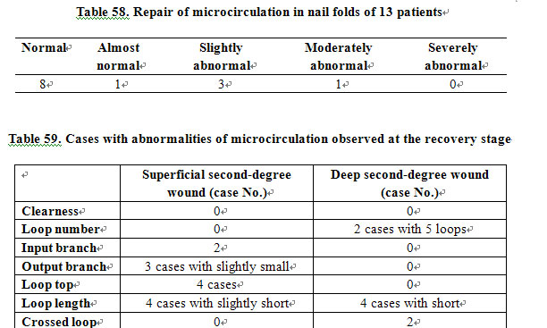
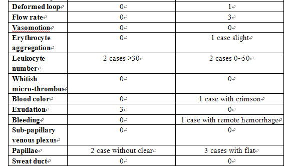
Changes in the appearance of the microcirculation of 6 patients with deep second-degree burns wounds were significant. Most of the loops were shorter. Two cases had 100% crossed loops. One case had a deformed loop exceeding the normal ratio. As a whole, parts of the microvessels in the nail fold resembled those in the superficial layer of the skin. Some cases had slower flow rate, which may be due to the changes of the loop appearance, such as crossed and deformed loops. One case had remote hemorrhage. Three cases had flat papillae, which may be related to the damage of deep tissue.
This study revealed that the microcirculation was repaired after treatment of burns wounds. This repair can be attributed to local application of BRT with MEBT/MEBO and its systemic benefit. MEBO protected the burns wound from dehydration and helped avoid bandaging pressure. This was favorable to the repair of the microvessels.
The microcirculation in the normal hands of these burns patients was examined and nothing abnormal was found (data not shown). The effect of other diseases could be excluded.
REFERENCES
1. Tian N (ed): Microcirculation: Basic and clinical. Beijing, Medical Science Press of CPLA, 1986, pp 343-345.